

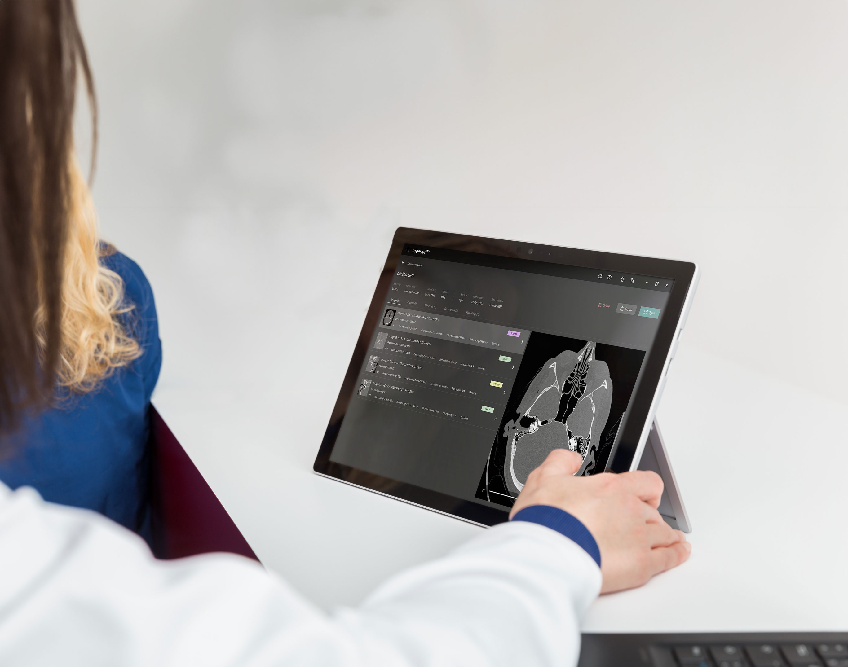
Data management
OTOPLAN® supports an automatic plug & play data import of DICOM files and allows to securely access imaging data from various sources like USB, external hard drive or PACS.
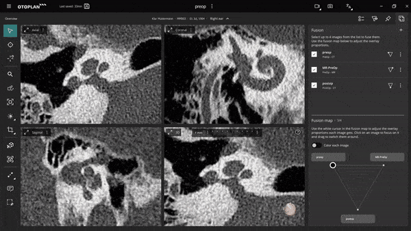
Fusion
Image fusion enables the user to see the best information in each imaging modality (CT and MRI) combined, or the pre-operative cochlear parameter measurements with the post-operative image for a faster and more consistent post-operative analysis.
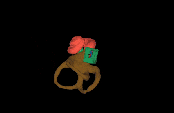
3D ear
Powerful 3D modelling algorithm reveals each patient's anatomy in full detail. This allows the user to measure delicate structures like the cochlea before the intervention and plan the best possible outcome for the patient by making full use of the patient's individual anatomical preconditions.
Cochlear implants
OTOPLAN facilitates accurate planning of cochlear interventions and is fully integrated into the HEARO system® for robotic cochlear interventions.
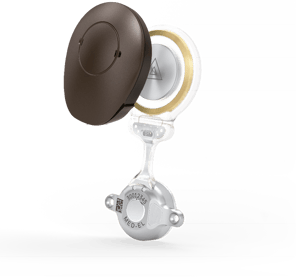
BONEBRIDGE®
It is possible to not only plan CI surgeries, but also other otological procedures. With OTOPLAN 3.0 you can also plan MED-EL’s bone conduction implant: BONEBRIDGE.
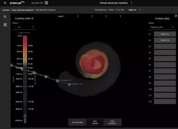
Electrode visualization
With OTOPLAN’s electrode visualization tool, you can compare electrode arrays from MED-EL’s comprehensive cochlear electrode array portfolio and see how each electrode array would match the unique cochlea of your patient. OTOPLAN uses individualized data to compare insertion depth and tonotopic pitch match with each MED-EL electrode array.
OTOPLAN makes it easy to discuss ideal electrode choice and individual surgical considerations with patients. One-step data export automatically generates a full case report in PDF or PowerPoint format for comprehensive consulting before and after the intervention.
Trajectory planning
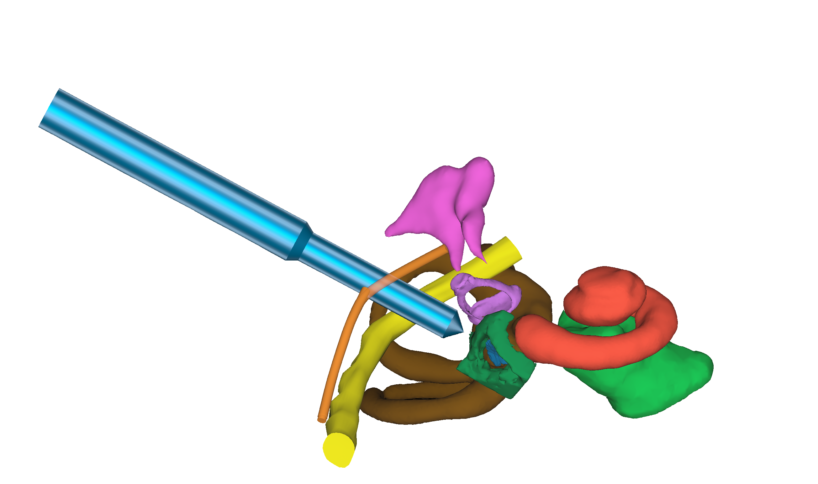
OTOPLAN helps aligning the insertion angle to the natural anatomy of the cochlea which can facilitate a smooth insertion of the electrode array into the scala tympani. At the same time the trajectory is planned with OTOPLAN in a way, that helps preserve critical structures like the facial nerve and the chorda tympani; represented in yellow and light orange respectively. This minimally invasive trajectory can be transferred onto the HEARO system to use it for robotic cochlear interventions.
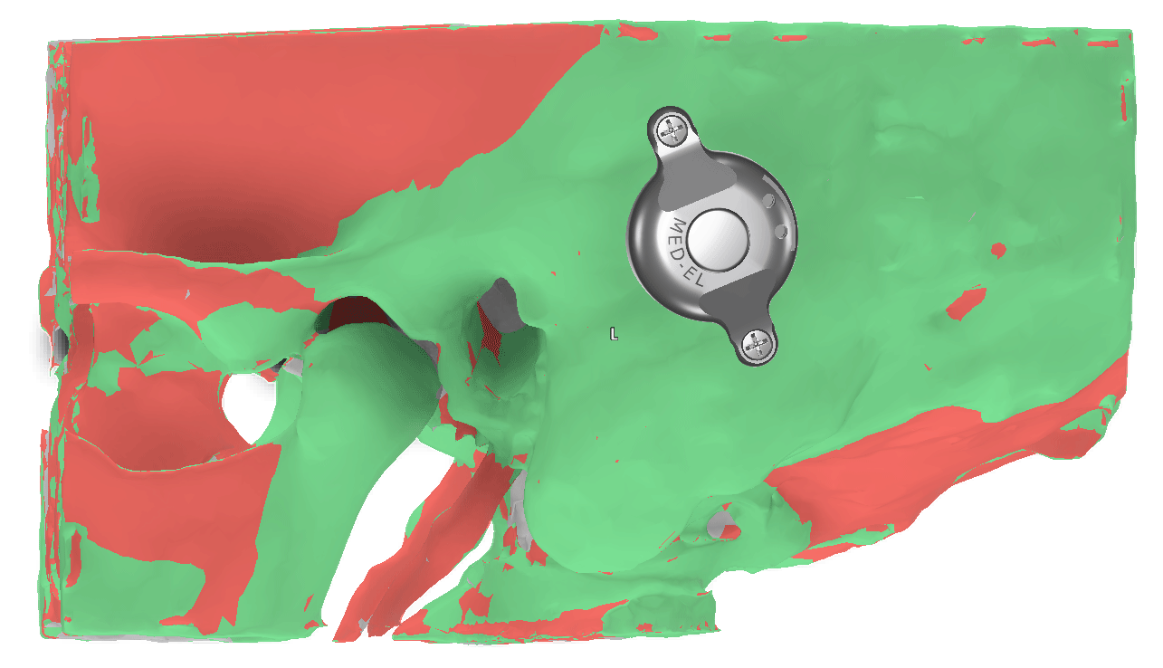
Thickness measurements
With the new automatic 3D reconstruction and thickness measurements of the skin and bone a custom heatmap can be created. This feature helps to visualize the optimal placement of the implant.
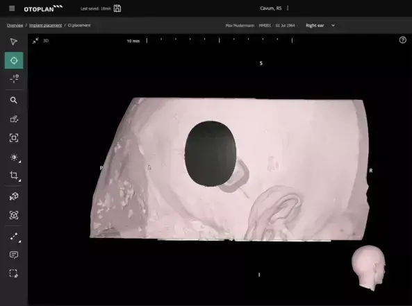
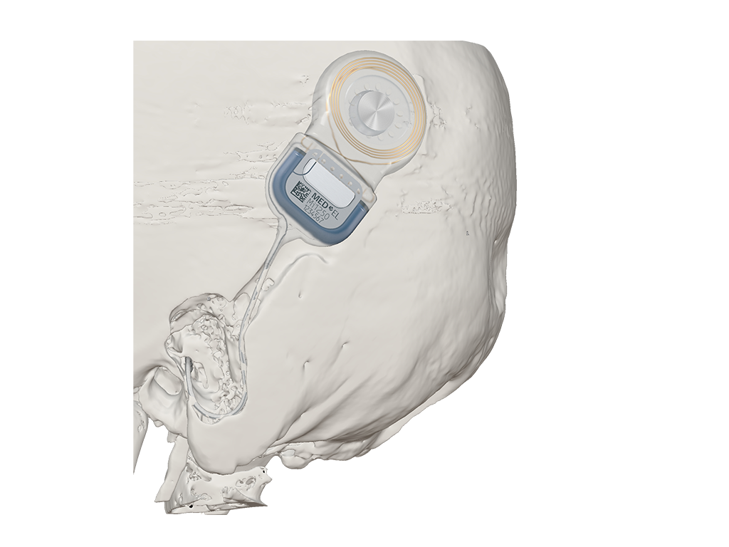
Placement planning
OTOPLAN supports uni- and bi-lateral placement planning for cochlear implants. Virtually move the implant housing and audio processor 3D models and easily mirror it to the opposite side. Together with the thickness measurements, this enables you to plan the placement in a safe and aesthetic location.
Implant detection
The latest version of OTOPLAN has also expanded its post-surgery assessment capabilities. The automatic implant detection is extended to also include the electrode lead and implant housing. This provides visual feedback on the insertion status and lead management.


| Previous version | Newest version | |
|---|---|---|
| Anatomy identification | ||
| Inner ear | Manual | Automatic |
| Temporal bone | Manual | Automatic |
| Skin | ||
| Applications | ||
| Cochlear implantation | ||
| BONEBRIDGE implementation | ||
| Audiograms | 2D | 2D and 3D |
| CI planning functionality | ||
| Cochlear parameters | Manual | Automatic |
| Virtual electrode insertion | 2D | 2D and 3D |
| Housing and processor placement (uni- and bilateral) | ||
| CI post-operative analysis | ||
| Detection of electrode contacts | ||
| Detection of electrode lead | ||
| Detection of implant housing | ||
| BONEBRIDGE planning functionality | ||
| Placement planning | ||
| Virtual cavity milling | ||
| General functionality | ||
| Multi-image data set management | ||
| Multi-image infusion | ||
| 2D and 3D measurement rulers |
1 |
5 (angular, linear, spline, polygon and freeform) |
| Multi-language | ||
| License manager |
Exclusively distributed by 
If you have any questions or need help, please reach out to MED-EL’s clinical support.

HEARO is the world’s first robotic surgical system for high precision, minimally invasive cochlear implantation.
By creating a tiny channel from the surface of the skull to the round window of the cochlea for insertion of the implant, it reduces trauma for the patient and enables to achieve consistent surgical results.